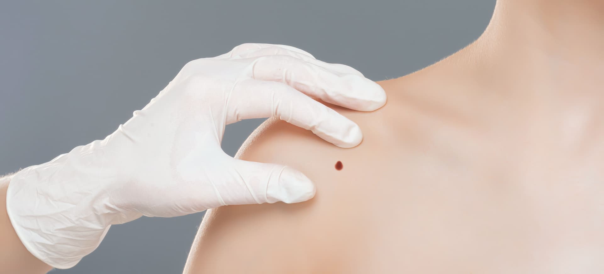
Cherry Angioma Removal in Irvine, CA — by Sabeen Munib M.D.
Diagnosis-first evaluation, then Nordlys IPL or electrocautery — never a quick zap.
Cherry Angioma Removal in Irvine, Orange County, CA
Cherry angiomas are the small bright red or purple bumps that appear most often after age thirty, usually on the trunk, arms, and shoulders. They are almost always harmless, but the only way to know that any individual lesion is harmless is to look at it first. At Spectrum Skin Clinic, removal is preceded by a focused diagnostic examination. Every red lesion is evaluated against the patterns that mimic cherry angioma before any cosmetic treatment is offered.
Dr. Sabeen Munib, MD selects between Nordlys IPL, electrocautery, or referral on the basis of lesion depth, surrounding skin, Fitzpatrick category, and what mimicker patterns may be present. For most patients with classic cherry angiomas, a single in-office session resolves the visible lesion with minimal downtime.

Spectrum at a glance
| Starting price | Google rating | Patient reviews | Physician-performed | Experience |
|---|---|---|---|---|
| Pricing shared at consultation | 5.0★ Google | 441+ across platforms | 100% by Dr. Sabeen Munib, MD | 15+ years |
Spectrum Skin Clinic — Irvine
114 Pacifica, Suite 280, Irvine, CA 92618 · (949) 259-4790
What are cherry angiomas?
Cherry angiomas, sometimes called Campbell de Morgan spots or senile angiomas, are small clusters of dilated capillaries in the upper dermis. They appear as bright red or purple papules, usually under five millimeters across, flat or slightly raised, with a smooth surface that blanches partially when pressed.
They are the most common acquired vascular lesion in adults. Onset is typically in the thirties and forties, with new lesions continuing to appear over decades. Hormonal shifts (pregnancy, perimenopause), genetic predisposition, and sun exposure have all been associated, though no single trigger explains every case.
Why the diagnostic step comes before treatment
Several lesions can look like a cherry angioma at first glance: amelanotic or nodular melanoma, pyogenic granuloma, angiokeratoma, spider angioma, and certain Kaposi sarcoma lesions. Dermoscopy at the consultation shows the vascular architecture characteristic of cherry angiomas, homogeneous red lacunae, and distinguishes most mimickers immediately.
When the appearance is atypical, the lesion is referred for biopsy rather than treated cosmetically. Treating a misdiagnosed melanoma with IPL or electrocautery delays the diagnosis and can compromise the histology. The diagnostic step is non-negotiable, which is why same-visit treatment is not offered on lesions that look unusual.

Treatment options at Spectrum
| Lesion pattern | Typical first-line choice | What it depends on |
|---|---|---|
| Multiple superficial cherry angiomas across face or chest | Nordlys IPL targeting the cluster | Skin type, lesion density, prior pigmentation history |
| Single raised or pedunculated cherry angioma | Electrocautery with light shave | Lesion thickness, proximity to mucosa or eyelid |
| Cherry angioma on Fitzpatrick IV–VI skin | Nordlys IPL with test spot first | Photoprotection habits, prior laser history, current inflammation |
| Rapidly growing, bleeding, or asymmetric red lesion | Biopsy and dermatopathology referral | Lesion features, change over weeks, systemic signs |
| Stable cherry angioma the patient prefers to leave alone | Periodic monitoring, no treatment | Patient preference, lesion stability, cost considerations |

Are cherry angiomas dangerous?
Most cherry angiomas are benign and never become malignant. They are not contagious and do not signal an underlying disease in the typical case. Lesions that are stable in size, color, and shape over months or years are reassuring.
What changes the calculus is sudden change. A lesion that grew quickly, bleeds without trauma, looks asymmetric, has variegated color, or is darker than typical red triggers a different evaluation. Those patterns are assessed as possible melanoma, pyogenic granuloma, or angiokeratoma, not treated as a cherry angioma. The first job of the consultation is making that distinction.

Electrocautery and shave excision as alternatives
For thicker or pedunculated cherry angiomas, electrocautery delivers a small controlled current that coagulates the lesion in seconds. A light shave can be combined when the lesion is raised enough to benefit from physical removal. Topical anesthetic is applied first; for larger lesions a small amount of local anesthetic is injected.
Healing forms a small crust over four to seven days. In Fitzpatrick IV–VI skin, electrocautery carries higher risk of post-inflammatory hyperpigmentation than Nordlys IPL, so the choice is biased toward IPL in melanin-rich skin when the lesion shape allows it.

Cherry angiomas in Fitzpatrick IV–VI skin
Cherry angiomas in deeper skin tones carry a higher risk of post-inflammatory hyperpigmentation around the treated area when settings are not adjusted. Spectrum's standard practice in Fitzpatrick IV–VI is a test spot before any first treatment, with lower starting fluence. The preference is IPL or shorter pulse durations over heat-based or sustained-energy approaches.
Patients who have had previous laser treatment elsewhere with rebound pigmentation are evaluated more conservatively. The plan may defer treatment entirely, or begin with a strict topical pigment-inhibitor regimen for several weeks before any device work.
Nordlys IPL for cherry angiomas: how it works
Nordlys is a broadband intense pulsed light platform with vascular-targeted filters. For cherry angiomas, a narrow wavelength range is selected so the energy is preferentially absorbed by hemoglobin inside the dilated capillaries. The vessels collapse and reabsorb over days to weeks, leaving the surrounding skin intact.
Most patients clear the treated lesions in a single session, occasionally two for larger or deeper angiomas. Sensation during treatment is brief, a series of warm snaps comparable to a rubber band, and no injectable anesthetic is typically required.

What to expect during the procedure
Diagnostic step: Each lesion is examined dermoscopically and photographed before any treatment begins.
Topical numbing: A short topical anesthetic is applied when patients prefer it; no needles for IPL.
Treatment time: Five to twenty minutes total, depending on lesion count and surface area covered.
Immediate appearance: Treated lesions may look darker, purple, or grey for a few hours before fading.
Same-day activity: Nordlys leaves the skin barrier intact; routine activities resume the same day.
Electrocautery healing: A small scab forms over the treated site and is left undisturbed for four to seven days.
Aftercare and downtime
Daily SPF: Broad-spectrum mineral sunscreen on the treated area for two weeks minimum, UV can trigger post-inflammatory pigmentation.
Avoid heat: No hot showers, saunas, or flushing-inducing exercise for several days.
Skip exfoliants: Alcohol-based products, acids, retinoids, and physical scrubs are paused until the skin is settled.
Leave crusts alone: Electrocautery crusts are not picked, scrubbed, or accelerated, they fall off on their own at day four to seven.
Return to routine: Most patients resume their full skincare regimen within seven to ten days.
Follow-up check: A four-to-six-week review confirms clearance and screens any new lesions of concern.
When a new red spot should be evaluated urgently
Most new red spots that look and behave like cherry angiomas can be evaluated at a routine consultation. The patterns that warrant earlier evaluation include rapid growth over weeks rather than months, and spontaneous bleeding that recurs. Color that is darker than red or contains brown or black, asymmetry, irregular borders, or association with systemic symptoms also shift the urgency.
These features do not confirm cancer, most still turn out to be benign, but they shift the next step from cosmetic treatment to diagnostic biopsy. Spectrum refers out for biopsy and dermatopathology when clinical examination warrants it, and cosmetic treatment is deferred until the diagnosis is clear.
Related Treatments Spectrum Does Offer
Cherry angioma removal often coincides with concerns about other small benign growths or vascular changes. Wart and skin tag removal addresses the other common acquired benign lesions Dr. Sabeen Munib evaluates at the same consultation. Vein removal is the appropriate option when facial spider veins or broken capillaries are present alongside cherry angiomas.
For diffuse facial redness or rosacea-type symptoms, laser genesis offers a gentler vascular-targeting approach in selected patients. To plan a combined visit that addresses cherry angiomas and adjacent concerns in one session, schedule a consultation with Dr. Sabeen Munib.
Red Marks That Are Not Cherry Angiomas
Not every small red mark on the face is a cherry angioma. Fine red lines that look like tiny branching threads across the nose or cheeks are usually broken capillaries, which are dilated vessels rather than a benign growth. They are treated differently. What causes broken capillaries on the face explains how to tell them apart and which light-based options apply. Where redness is diffuse rather than a discrete spot, rosacea treatment is usually the more relevant page.
Book a cherry angioma consultation in Irvine
Spectrum Skin Clinic in Irvine, California sees patients from across Orange County and Los Angeles for cherry angioma evaluation and removal. The consultation includes dermoscopy of the lesions of concern, recommendation of treatment modality (or no treatment) per lesion, and a written plan with realistic timelines.
Patient Reviews
4.97★ · 441 Patient Reviews

I had an amazing experience with Dr. Munib, who performed my aesthetic treatments including Pico laser, Botox, a glassy facial, Botox for migraine, and HIFU. Every procedure was done with great care, precision, and professionalism, and the results exceeded my expectations. Dr. Munib is incredibly knowledgeable and takes the time to explain everything, making me feel comfortable and confident throughout the process. Her expertise truly shows in her work. The staff was extremely accommodating, friendly, and professional, which made the entire experience even better. On top of that, the prices were very reasonable for the quality of service provided. I highly recommend Dr. Munib to anyone looking for outstanding aesthetic treatments and excellent patient
Mahjabeen Saeed
4 months ago

I had an excellent experience with Dr. Sabeen Munib at Spectrum Skin Clinic in Irvine. I visited her for acne scar treatment and underwent three subcision sessions spaced six weeks apart. Throughout my treatment, I was consistently impressed by her expertise, attention to detail, and compassionate approach. Dr. Munib took the time to carefully evaluate my scars, explain the procedure, and answer all of my questions before starting. During each procedure, she made sure I was comfortable and informed every step of the way. Her technique, skill and experience with acne scar subcision gave me complete confidence that I was in capable hands. The entire team at Spectrum Skin Clinic was professional, friendly, and supportive throughout the process. Although acne scar improvement takes time and healing, I appreciate that Dr. Munib focuses on achieving the best possible long-term results rather than making unrealistic promises. She explained what to expect during recovery and emphasized that collagen remodeling happens gradually. If you're looking for a knowledgeable and caring physician for acne scar treatment or subcision, I highly recommend Dr. Sabeen Munib Spectrum Skin Clinic, Irvine. I truly appreciate the care, professionalism, and dedication I received and would not hesitate to recommend her to anyone seeking advanced scar treatment.
Deepak Jadhav
3 months ago

After several aesthetic nurses told me I was allergic to filler dissolver, Dr. Munib took a careful, evidence-based approach. She used ultrasound to locate my migrated lip filler, tested a small area first, and explained I was reactive—not allergic. She dissolved a small test area before safely treating my upper lip. Throughout the process she was gentle, thorough, and educational. It’s a long drive for me, but her knowledge and expertise make it absolutely worth it.
Jamel Neill
2 months ago

Thank GOD for Dr. Munib. She is the angel of dissolver!!! I had HA nodules and Sculptra nodules from previously going to Dermfx and I went to Dr. Munib for ultrasound guided dissolving. She is gentle, patient, knowledgeable, kind and funny. I feel like she is the last of the good ones in this business. If you are interested in natural looking aesthetic results that aren’t botched and don’t cause you years of facial pain go to her. I would try to see her as my primary doc if I wasn’t moving out of the area. I would travel to go see her though.
Siena Viencek-Walsh
3 months ago

Frequently Asked Questions
Got questions? We’re here to help! Find out what we offer, how to book an appointment, where we’re located, and more. We treat all age groups and offer skincare products too.